Many terms are used to describe issues with a spinal disc and disc pain, and they are often used differently and, at times, interchangeably. Some commonly used terms include:
- Herniated disc
- Pinched nerve
- Ruptured or torn disc
- Bulging disc
- Disc protrusion
- Slipped disc
There is no firm consensus on the use of these terms, and as a patient it can be frustrating to hear one diagnosis described in so many different ways.
Watch: Herniated Disc patient education
Two Causes of Pain: Pinched Nerve vs Disc Pain
There are two main ways a spinal disc can cause pain:
- Pinched nerve. In most cases a herniated disc itself is not painful. Rather, the material leaking out of the disc pinches, inflames or irritates a nearby nerve, causing radicular pain. Radicular pain (also called nerve root pain) describes the sharp, shooting pains that radiate to other parts of the body, such as from the low back down the leg or from the neck down the arm. Leg pain from a pinched nerve is commonly called sciatica.
- Disc pain. A spinal disc itself may be the source of pain if it dehydrates or degenerates to the point of causing pain and instability in the spinal segment (called degenerative disc disease). Degenerative disc pain tends to involve a chronic, low-level ache around the disc, with occasional episodes of more severe pain.
A herniated disc and degenerative disc disease typically occur in the cervical spine (neck) and lumbar spine (lower back). Disc pain tends to be most common in the lower back, where most of the movement and weight-bearing in the spine occurs. These conditions are uncommon in the mid-back (the thoracic spine).
Diagnosing Disc Problems
Review of Medical History and Specific Symptoms
The diagnostic process typically begins with a review of your medical history and current symptoms. A complete review of symptoms will include:
- The location of the pain, including whether it is confined to the neck or back, or whether it includes arm or leg pain
- A description of how the pain feels, such as searing, sharp or stabbing, compared with dull or achy
- Whether certain activities, positions or treatments make the pain feel better or worse
Collecting a full medical background can rule out or identify other possible conditions that may cause pain. A medical history may include information on recurring health problems, previous diagnoses, and past treatments and their effectiveness. Information on sleep, diet and exercise habits is usually also collected.
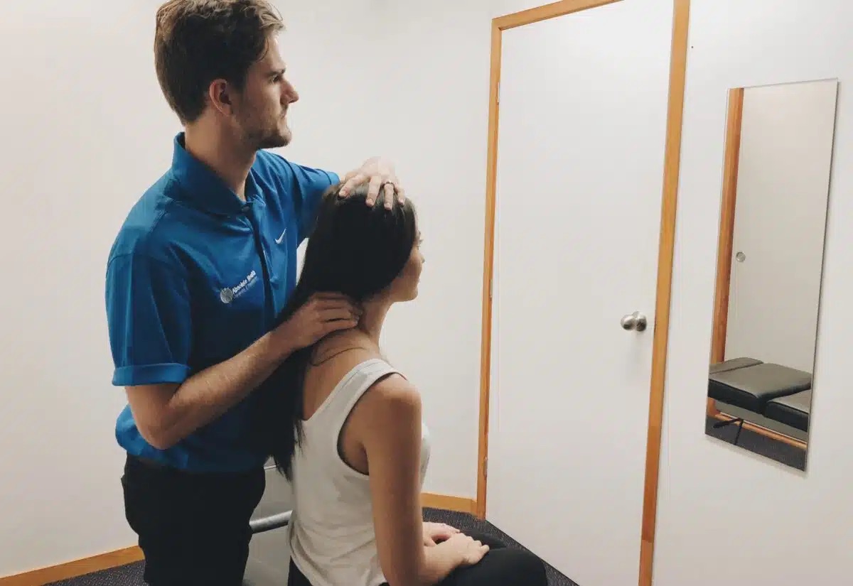
Physical Examination
A physical exam for diagnosing disc pain may include one or more of the following tests:
- Palpation. Palpating (feeling by hand) certain structures can help identify the source of pain. For example, worsened pain when pressure is applied to the spine may indicate sensitivity caused by a damaged disc.
- Movement tests. Tests that assess the spine's range of motion may include bending the neck or torso forward, backward or to the side. If raising one leg in front of the body worsens leg pain, it can indicate a lumbar herniated disc (the straight leg raise test).
- Muscle strength. A neurological exam may be carried out to assess muscle strength and determine whether a nerve root is compressed by a herniated disc. A muscle strength test may include holding the arms or legs out to the side or front of the body to check for tremors, muscle atrophy or other abnormal movements.
- Reflex test. Nerve root irritation can dampen reflexes in the arms or legs. A reflex test involves tapping specific areas with a reflex hammer. If there is little or no reaction, it may indicate a compressed nerve root in the spine.
Some physical exam tests may be used to rule out or confirm a diagnosis that coincides with symptoms reported in the medical history.
Diagnostic Tests
A diagnostic test may be ordered to confirm the disc problem and to gain additional information, such as the location of a herniated disc and any impinged nerve roots. Diagnostic tests may include:
- CT scan / myelogram. Computerised tomography (CT) scans consist of an X-ray of the body, with a computer reformatting the image into cross sections of the spine. Sometimes a myelogram is performed during a CT scan, in which radiographic dye is injected into the area to provide more detail on the spinal structures. See Computerised Tomography (CT Scan).
- MRI scan. Magnetic resonance imaging (MRI) provides a sensitive and accurate assessment of the spinal nerves and anatomy, including disc alignment, height, hydration and configuration. See MRI Scan of the Spine.
- Discogram. A discogram may be recommended to confirm which disc is painful if surgical treatment is being considered. In this test, radiographic dye is injected into the disc to recreate disc pain through the added pressure. See Discogram.
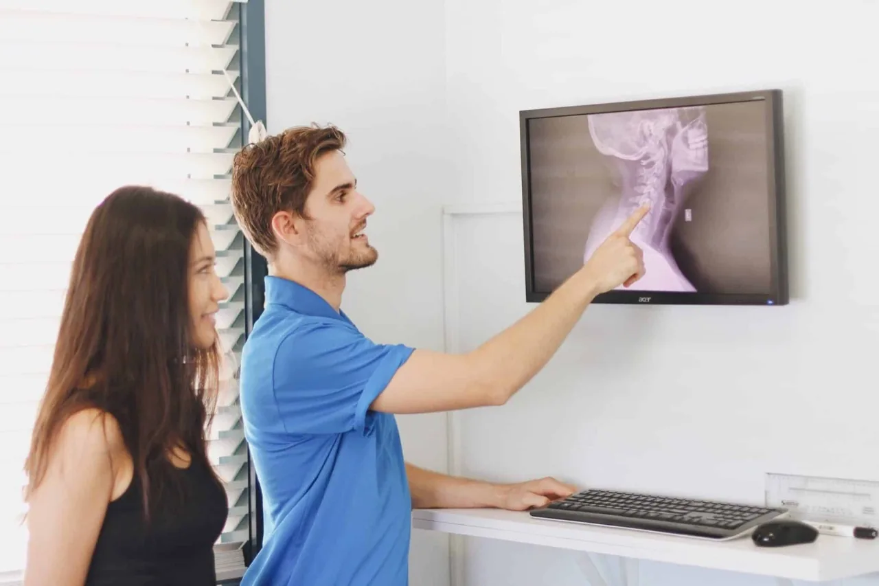
- X-rays. Although plain film X-ray is a poor imaging method for visualising the disc itself, it can provide important feedback about what is causing the disc problem and the state of the disc.
It is important to note that the findings on an MRI scan or other diagnostic test are not in and of themselves a diagnosis. Physical exam findings and a review of symptoms need to match the MRI or other test findings to accurately identify the cause of pain.
Only then can an effective treatment plan be prescribed, whether that involves treating a pinched nerve from a herniated disc, disc pain from degenerative disc disease, or another condition.
At Absolute Health we have two clinics on the Sunshine Coast, in Mooloolaba and Nambour, where we treat disc conditions all the time. If you are suffering from this condition, contact our office to see if we can help today.